5 Tools to Diagnose Impacted Teeth
- CephX | AI Driven Dental Services
- Blog
- 5 Tools to Diagnose Impacted Teeth
5 Best Tools to Diagnose Impacted Teeth
Misdiagnosed impacted teeth may turn out to be risky to dentists just s they are to patients. Malpractice lawsuits are increasing in numbers, as patients claim for negligence- failure to timely recognize the need for eruption management, and failure to take timely action before risking the patients’ health.
The causes of an impacted tooth are often as a result of lack of space for the tooth in the mouth, due to orthodontic treatment or genetic factors. In other words, it may be caused by inherited factors or as a secondary effect to another oral condition.
This tooth condition can present with symptoms, i.e., symptomatic or even show no symptoms at all, i.e., asymptomatic. For those that present with symptoms, the common ones are:
- Bad breath
- Red swollen gum
- Pain associated with mouth opening, biting, or chewing.
- Bleeding gum etc.
The list of symptoms is not exclusive; some may experience one or more, while others none. And of course, these symptoms can serve as valid hallmarks to diagnosing the impacted tooth condition.
Here are some of the best methods of diagnosing impacted teeth:
- Evaluation of the condition of tooth and gum: This is the conventional first diagnostic step taken by dentists. But this diagnosis is often not accurate, as there are many gums, teeth, or oral disease conditions that affect the gum, presenting almost the same symptoms as the “red- swollen- bleeding gum.” However, it can give a solid lead.
- Intra-oral radiography – This type of radiography is said to more commonly used compared to the extra-oral radiography. The various Subtypes under it are indicative of the aspect of the teeth they show. Such Subtypes include: Bite-wing x-rays, Periapical x-rays, Occlusal x-rays.
Known down-side for intra-oral radiography is their limited field-of-view, which can easily lead to missing out impacted teeth even when pointing the equipment to the right area.
- Extraoral radiography – Referring primarily to Panoramic x-rays, this is a method preferred by most dentists. It enables effectively detecting teeth impaction within a region of the mouth. This implies that the specificity of location of impacted teeth may not be ascertained.
- Cone-Beam Computed Tomography (CBCT): Acquiring 3D scan is usually better, safer and more accurate than the radiography methods. For the safety of the patient it is often safer to take one exact CBCT in place of having to do repetitive radiography checks. The harmful effect of radiation is thus circumvented by this method of diagnosis. However many dentists find it hard to master 3D software, having to search through slices using different filters. Also, unlocking its full potential requires manual anatomy segmentation, before teeth and jaws can be moved, colored, removed etc, to allow diagnosis of impaction and other clinical condition, such as root absorption, bone thickness, nerve canal location and others.
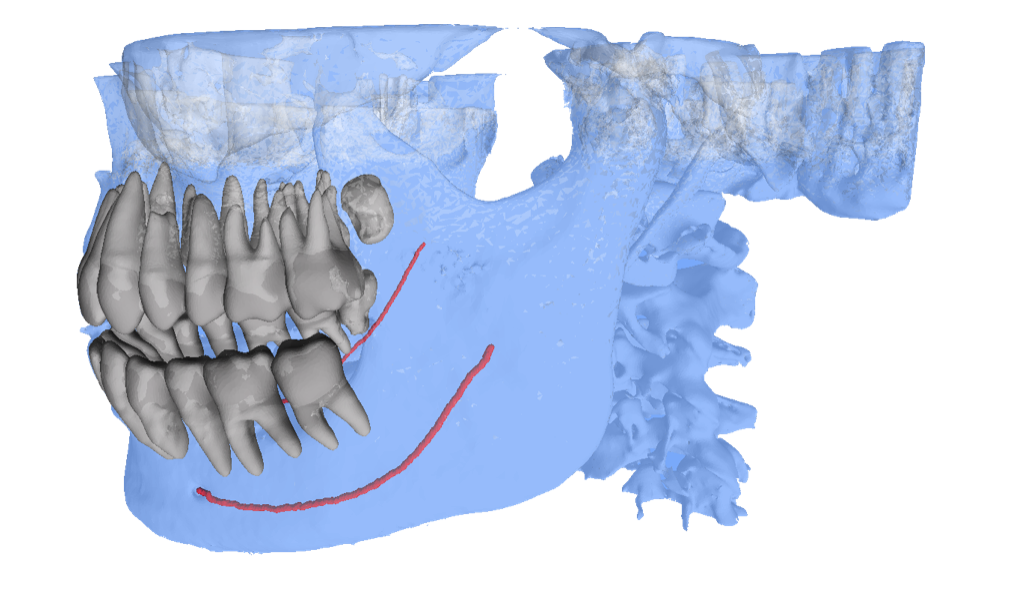
- AI Driven 3D Segmentation: A 3D viewer that shows teeth and bone segmented by artificial-intelligence from a CBCT scan. This is an improvement over the conventional cone-beam computed tomography method of diagnosis, without adding any manual segmentation work or cost

Unlike the other methods of diagnosing impacted teeth, this newer method is better and more reliable. It gives a perfect 3D picture of the teeth segmented, making it very easy for the dentist/oral surgeon to accurately identify the impacted tooth/teeth. Dentists also have the option to share the case with patients, making them more connected and informed, to increase case acceptance.
CBCT 3D viewer helps the dentist to precisely identify the location of the impacted teeth; this is unlike the panoramic imaging that only helps to predict the teeth such as the maxillary canine. It reveals the location and shows the entire teeth in a three-dimensional manner, thus enabling accurate diagnosis.


