Dr. Zeev Abraham (DMD, MS, MSc)
This example case demonstrates the value of new digital technologies, when using the cephX (Orca) automated teeth segmentation service for CBCT data.
Patient History:
Patient Profile: Patient is 13-years old male.
Classification: Skeletal Class l Medium crowding in Maxilla Mild crowding in the mandible Bilateral Crossbite
Dental History: No restorations (only Fissure Sealant) ; A few prior visits to the dentist.
Breathing: Combined mouth and nose.
Case Evaluation:
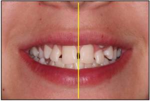
Symmetric soft tissue smile
Upper midline coincides with mid-facial line
Exposure of 95% of central incisors
Slight gum exposure
Lower teeth shown in smile
Lower midline 2 mm right of uppermidline (dental shift)
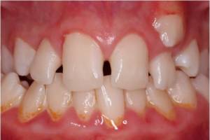
Crossbite – 14 to 17, and 24 to 35
Tooth 13 unerupted and impacted
Tooth 23 ectopic and eruptedbuccal to 22
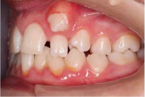
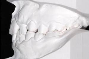
Intraoral Evaluation vs. Study Model (Left)
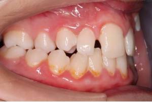
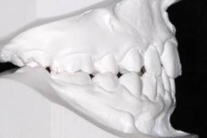
Intraoral Evaluation vs. Study Model (right)
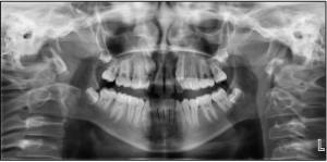
Panoramic Analysis:
The Condyles are symmetrical
3rd molar buds developing
Tooth 13 is impacted
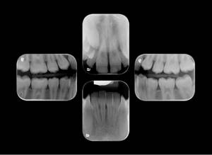
The root of 12 is probably resorbedPeriapicalsX-Rays:
 Dilacerations of lower incisor roots
Impacted Tooth 13 partially buccal and partially palatal to 12 root.
Impacted 13
Dilacerations of lower incisor roots
Impacted Tooth 13 partially buccal and partially palatal to 12 root.
Impacted 13
 CBCT
CBCT
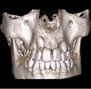
Discussion:
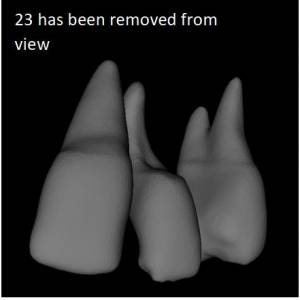
In the panorex, generated by the DICOM of a standard CBCT, there is a suspicion that the root of 12 has been resorbed by the impacted 13 situated above and palatal to its root. The extent of resorbtion is not clear in any of the relevant anterior/posterior cuts numbered 12 to 16. There is no suspicion that the root of 22 has been damaged by the buccaly positioned 23, to the extent that the technician did not even identify any 2D transverse cuts in that area. However, in the 3D STL file generated by the cephX’s automated segmentation algorithm, we could clearly see the relationship and extent of resorbtion of the 12 and 22 roots. Video – https://youtu.be/Y_2r6Hhgo8o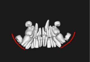 A video of the segmentation, generated automatically by CephX
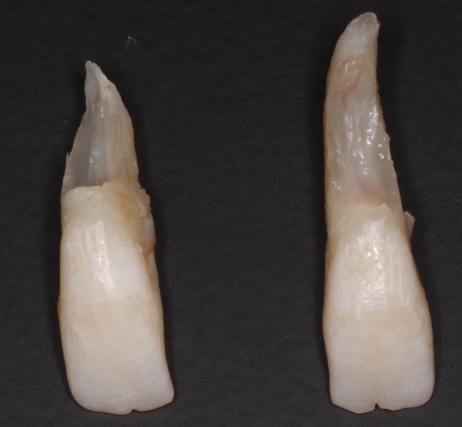
Of particular interest, the 22 root was unsuspectedly and uncharacteristically resorbed on its buccal side while leaving the absolute length of the root intact.
A video of the segmentation, generated automatically by CephX
Of particular interest, the 22 root was unsuspectedly and uncharacteristically resorbed on its buccal side while leaving the absolute length of the root intact.
Conclusion:
As a result of the clear resorbtion of 12 and 22 roots, there is a valid reason to extract both the 12 and 22 and resolve space requirements accordingly CephX software was a tremendous help enabling quick detection and diagnosis of the resorbed teeth. The STL files that were generated by CephX AI allowed us to view each tooth separately and to have a better understanding of the patients’ intra-alveolar dental layout.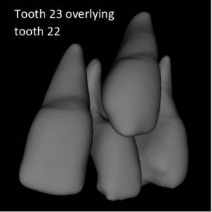
After the upper laterals were physically extracted, the actual anatomical resorbtion of both lateral roots could be compared to the STL view of the exposed laterals. The amount and position of the root deformation was remarkably similar when comparing the STL exposed image view and the actual teeth themselves. The segmented STL view of the teeth and jaws is a new powerful tool in the diagnosis and treatment planning of impacted teeth, resorbed roots, mandibular nerve relationships and alveolar bone thickens.