Interest in measuring the human form and skull has been around for millennia. The history of
cephalometric analysis stems from Egyptian and Greek attempts at human body measurement (anthropometrics).
The term cephalometric is sometimes confused with craniometric. The former refers to measurement of the skull. The latter refers to measurement of the head including the soft tissue be it living or dead.
History of Cephalometric Analysis/Pre-Roentgen
4000 (BC) Egyptians: Canon of Proportions was a mathematical system developed to give idealistic proportions to the human form. Artistic forms were generated using a grid system.
Evidence of attempts at orthodontics and tooth “bridging systems” using wires have been found among the human remains of ancient civilizations. The idea that modern day orthodontics may have originated thousands of years ago, is certainly intriguing. This remaining ancient physical evidence of attempts at orthodontics indicates that the dentition and facial appearance were as important then as they are today.
* Although not strictly related to cephalometric contributions, attempts down through time have been made to qualify and quantify the human form by type. *
- (c.460-c.370) Hippocrates: described two body types.
Habitus phithicus: Long thin body
Habitus apoplecticus: Short thick body
- (1921) Kretschmer:
Pyknic: Fat and stocky
Asthenic: Weak, small and thin
Athletic: Muscular and large boned
- (1954) Sheldon:
Endomorph: tending toward body fat
Mesomorph: tending toward musculature
Ectomorph: tending toward undeveloped muscle
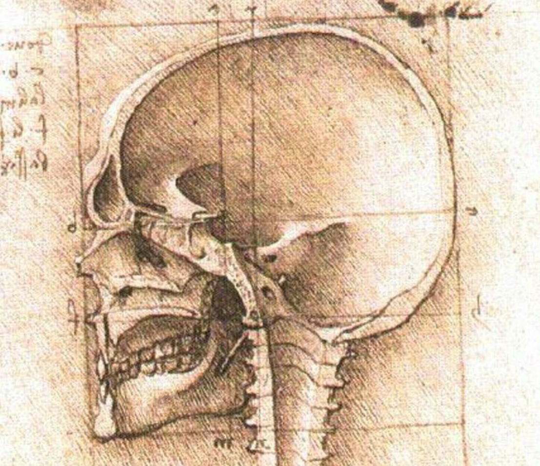
- (1452-1519) DaVinci: Arguably the first to try and systematically measure the head.
- (1528) Albrecht Durer: A treatise on cranial measurements was the first published work in which anthropometry was applied to aesthetics.
- (1678-1761) Pierre Fouchard: Published the “Surgeon Dentist” in 1728. Some consider him to be the “inventor” of orthodontics. Some of the “less than modern” methods of straightening teeth included finger pressure, metal plates lashed to abutment teeth, extractions and the use of a surgical instrument of the time called “The Pelican”. This instrument was used to make large forceful lingual to buccal tipping movements. If possible, it would have been interesting to talk to some of his teenage patients!
- (1722-1789) Petrus Camper: introduced the facial angle, facial line and horizontal plane.
- (1847) Joachim LaFoulon: Coined the term
- (1796-1860) Anders Retzius: Credited with introducing the terms orthognathic, prognostic and the cephalic index.
Kingsley and Farrar are credited with being the “Fathers of Orthodontics”. Both wrote definitive books on orthodontics during the late 1800s.
Edward Angle was a paramount figure in orthodontics. His work in the early part of the twentieth century remains an influence in present day orthodontics.
Roentgen Leads The Way
(1895) Roentgen discovered X-rays in 1895 and submitted the paper, “On a New kind of Rays, a Preliminary Communication”. The following year Koening and Walkhoff simultaneously made the first dental X-ray of a tooth.
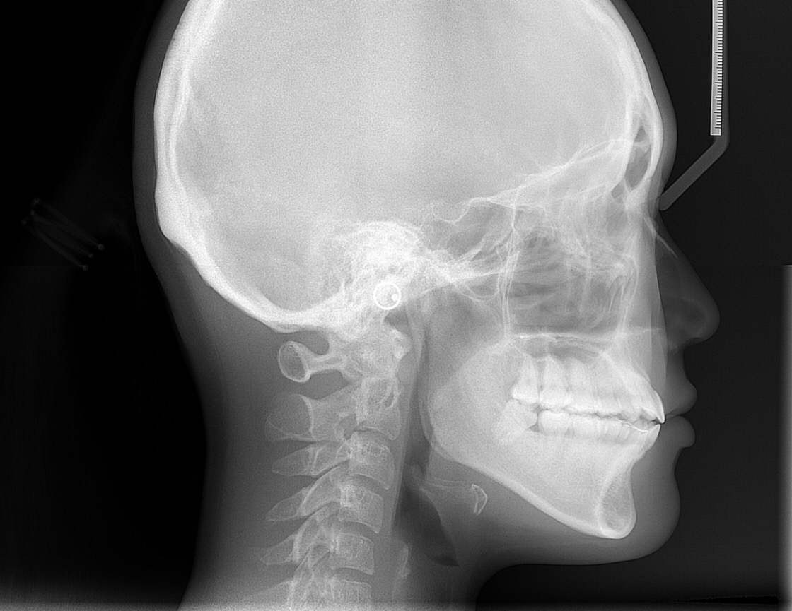
(1922) AJ Pacini is credited with making the first standard lateral view radiograph in 1922.
(1922) Paul Simon (Germany) becomes the first to use planes and angles in the diagnosis of dental anomalies
(1922-1931) During this period, various researchers reported on the use of radiographs in the practice of orthodontics. These contributions included the discovery of new radiographic landmarks and various attempts to incorporate and improve methods of diagnostic measurement including attempts to obtain a standardized practical method for obtaining radiographs.
(1931) Holly Broadbent along with Todd Wingate (United States), H. Hofrath (Germany) simultaneously developed the cephalostat. Broadbent occupies a special place in the evolution of cephalometrics as many of his principles and ideas have been accepted practice since their inception. Broadbent used a metric scale and reproducible head positioning of the cephalostat to eliminate the problems associated with previous unstandardized radiographic analysis.
(1937-1947) Much of the evolution of cephalometric analysis during this period was associated with investigating the craniofacial growth factors affecting orthodontic treatment (down and forward, Brodie 1941) and other factors relating to the dentation in its relationship to various craniofacial factors, (Margolis 1943, inclination of incisors).
(1948) William Downs is credited with developing the first cephalometric analysis. Over the ensuing years multiple cephalometric analysis methods have been established,
- (1953) Steiner
- (1954) Tweed
- (1955) Sassouni
- (1974) Harvold
- (1975) Wits
- (1979) Ricketts
- (1985) McNamara
- (1972) Jaraback
Technologies
Initially, cephalometric analysis was performed manually using acetate tracing paper and a lighted view box. Tracings of pertinent diagnostic lines using established orthodontic anatomical landmarks were drawn using a #3 lead pencil. This method of performing an analysis brings in problems of accurate analysis due to human error and differences of experience and expertise of the analyst.
Digital radiographs
(1960s) The proposition of digitalized radiographs as a vehicle for cephalometric analysis came into play and technologies have evolved since that time to make the process faster, easier and more accurate. Types of digital radiographs include:
- Indirect digital
- Direct
- Semi-direct
Computed Tomography (CT) MRI, PET, PET/CT
(1971) First used in the United Kingdom. Technology improvements included reduction in time for the procedure due to increased number of slices produced in the same rotational period and increases in image size. Other imaging modalities followed.
- (1980) Magnetic Resonance Imaging (MRI)
- (1985) Positron Emission Tomography (PET)
- (2000) Positron Emission Tomography/ Computed Tomography (PET/CT)
Initial dental use of CT was necessarily restricted due to the size and expense of the equipment. However, this technology was the basis and cornerstone of today’s use of CBCT in the science of cephalometric analysis.
Cone Beam Computed Tomography (CBCT)
(1995) Tacconi and Mazzo develop a system to utilize CBCT technology for dental purposes.
(2001) CBCT was first introduced in the United States after the “New Tom 9000” machine was approved by the FDA. Using a cone beam rather than a fan beam along with other innovations allowed the equipment for CBCT to be reduced in size enough to be installed in the average dental setting and at a (comparably) affordable price.
(2007) Kodak introduces Ultra CBCT ILUMA scanner
(2007-present) Competition has resulted in different brands of CBCT machines offering their own design, branding advantages, etc. Most often these differences are related to field of view (FOV) and improvements in image resolution.
The increased use of CBCT has resulted in the necessity of a parallel technology improvement in rendering cephalometric analyses. Technology has resulted in computerized software models for in-office and online cephalometric analysis.
Founded in 2001, the online company Cephx has been at the forefront of developments in CBCT cephalometric analysis rendering technology. These developments in technology allow Cephx to provide orthodontists and other dental practitioners with cutting edge solutions regarding analyses, cloud storage and patient records management.
For more information please contact
info@cephx.com or 1-800-992-1499