Why Patients Leave Before Treatment – And How DSOs Can Win Them Back
Dental Support Organizations (DSOs) are expanding rapidly, but one persistent challenge continues to erode both revenue and patient trust: patients leaving before starting, failing to complete treatment, or disengaging after just one consultation.”t. Research shows that nearly 6 in 10 patients fail to return after their first dental appointment – often due to confusion, financial concerns, or lack of trust ¹
So why do patients “ghost” before treatment completion or initiation- and more importantly, how can DSOs change the outcome?
The Patient Retention Challenge² ³
- Confusion and Overload
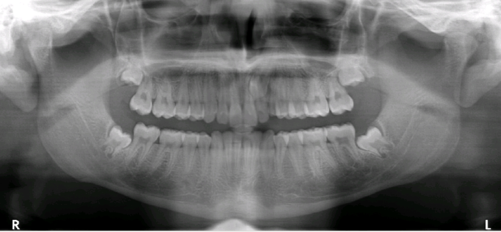
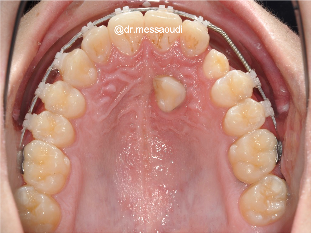
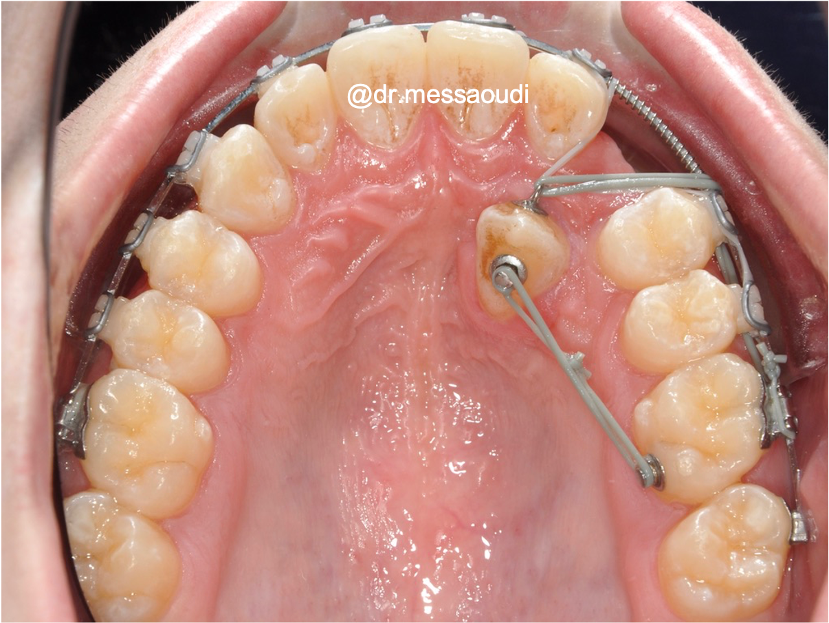
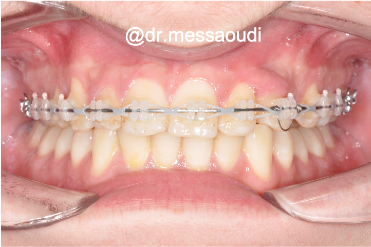
Dental treatment plans can be overwhelming. When explained only in clinical jargon or unclear imaging such as x-rays, many patients struggle to understand the diagnosis or necessity, leading to hesitation - Weak Case Presentation
Verbal explanations alone often fail to connect. Without visual reinforcement, patients are left uncertain about their condition and the recommended treatment. - Financial Anxiety
Unclear pricing or lack of financing options can scare patients away. Transparency is key to retention . - Lack of Engagement
When DSOs fail to follow up after consultations, patient interest drops off. Without proactive communication, momentum is lost.
What DSOs Need to Do Differently
The most successful DSOs are rethinking the patient journey:
- Using digital, visual treatment plans to bridge the understanding gap.
- Offering clear financial breakdowns and financing options.
- Leveraging AI-powered tools to build trust and improve efficiency.
- Maintaining consistent follow-up to keep patients engaged.
In fact, DSOs that prioritize the patient experience have seen case acceptance jump from around 43% to as high as 96%⁴.
How CephX Helps DSOs Convert Patients
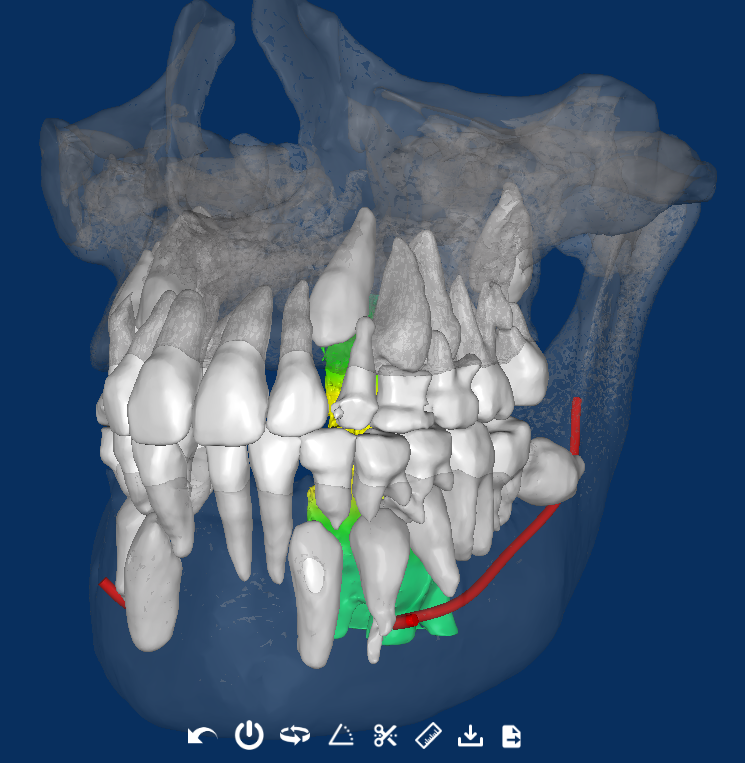
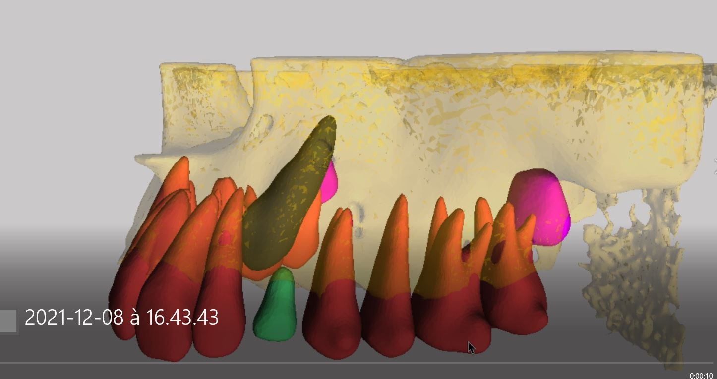
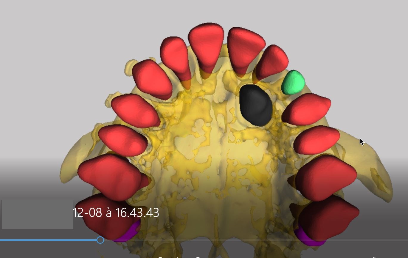
This is where CephX makes the difference. By combining AI-powered diagnostic tools and visualizations that can be easily conveyed to the patients, CephX empowers DSOs to present treatment journeys in ways that patients can understand and are able to trust.
- Visual Clarity That Builds Trust
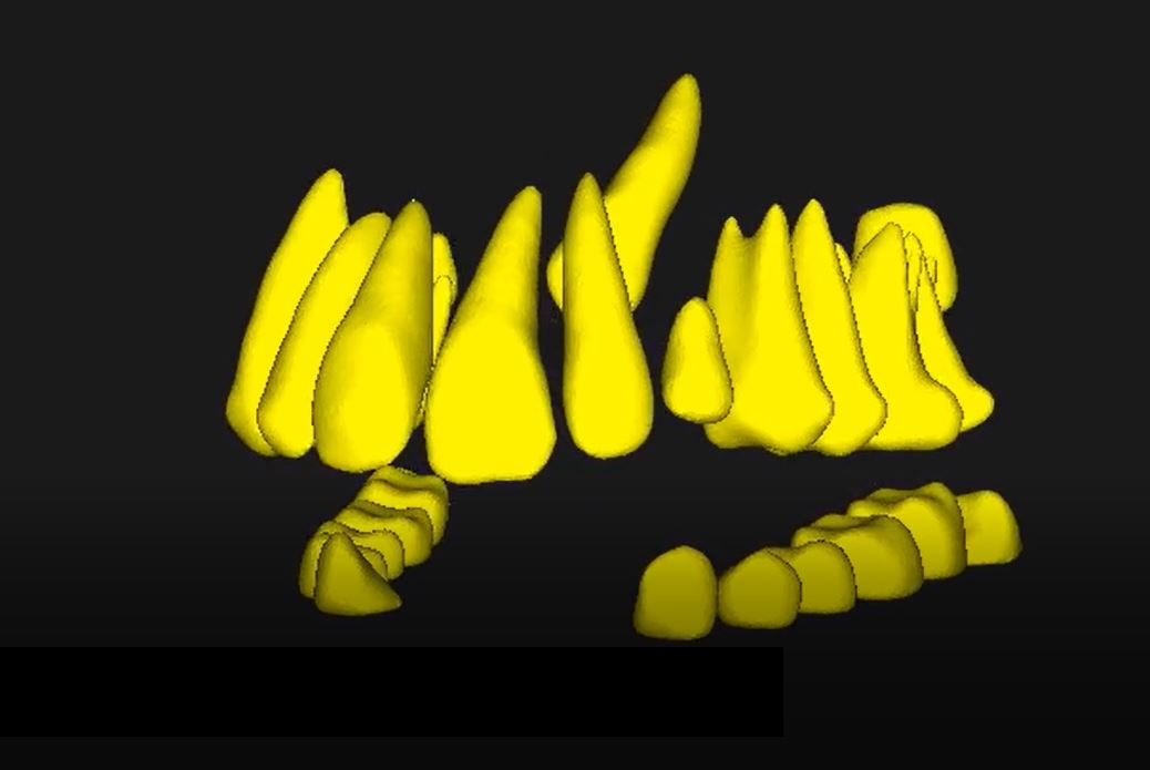
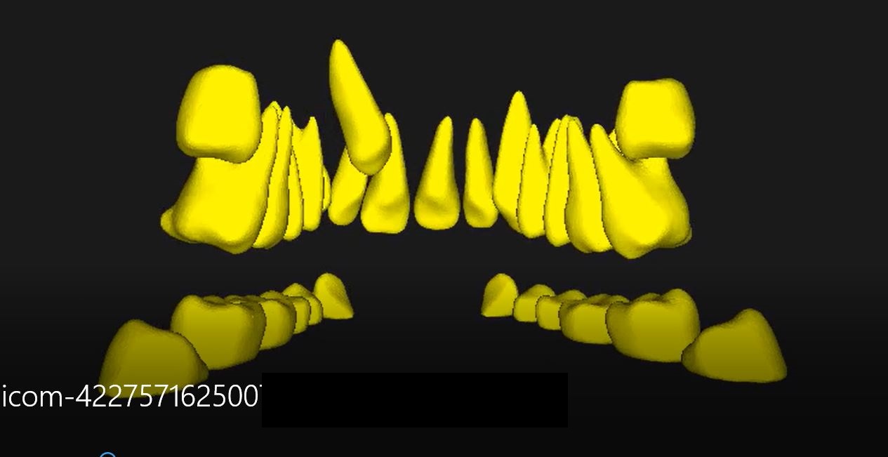
CephX automatically analyzes various case related images to produce visual explanations that allow patients to comprehend the clinical finding and the proposed solution - Efficiency and Precision
AI-driven automation replaces manual digital processing , delivering accurate results in minutes, so DSOs can present treatment journeys at chairside - Stronger Case Presentations
By presenting smart visuals , both of the clinical findings as well as the desired treatment outcome , patients feel an elevated level of confidence that the treatment suggestion is necessary,effective and safe. - FDA-Cleared Reliability
CephX’s AI platform has received FDA clearance, giving DSOs a clinically validated tool that patients can trust .
Conclusion: From Missed Opportunities to Completed Treatments
For DSOs, every patient who walks away before treatment represents lost revenue and lost opportunity to deliver care. The solution lies in better communication, visual engagement, and trust-building early in the patient journey.
By adopting AI-powered tools like CephX, DSOs can bridge the gap between diagnosis and decision, helping patients not only understand their treatment but feel confident enough to move forward.
Want to learn more >>>